Leishmaniasis is a disease caused by protozoan parasites, and is spread by sand-flies. There are currently three different types of the disease, cutaneous, mucocutaneous, and visceral leishmaniasis. Yes, those are some pretty long and serious sounding diseases, but the symptoms are the things that should be frightening you, which include skin sores, volcano-like ulcers, skin ulcers, mucosal ulcers, and damage to internal organs. That all sounds pleasant . . . not.
Earlier this month, the news media hyped a story that the Islamic State was causing the spread of leishmaniasis, because – as the U . K . ’s Mirror newspaper put it – militants were “slaughtering innocent people and dumping their bodies in the street.”
Leishmaniasis has been spreading like wildfire in Syria since the health system collapsed in rebel-held territories in 2011. By 2012, there were already 52,982 documented cases of the disease in Syria.
Also in 2012, the CDC documented that “migration patterns of refugees with cutaneous leishmanias is were identified in Lebanon,” with the health agency producing a helpful illustration showing the disease’s “movement from cities in Syria to regions in Lebanon.”
The peer-reviewed medical journal Pathogens noted that Lebanon had no cases of cutaneous leishmaniasis prior to 2008 and only “sporadic cases in the following years.”
After the arrival of refugees, 1,033 cases were confirmed by 2012, “96.6% (998) of which were among Syrian refugees.” Writing at AHC Media, a publication for healthcare professionals, Dr. Philip R. Fischer, Professor of Pediatrics at the Mayo Clinic, documented the spread to Turkey as well:
As Syrians leave their homeland, they sometimes carry their germs with them. There have been dramatic increases in the number of cases of cutaneous leishmaniasis in southeastern Turkey. In Turkey, 69% of cutaneous leishmaniasis patients are Syrians living in tent cities.
Fischer also noted a significant risk of the disease spreading to Europe with the arrival of Syrian refugees.
As recent news reports have shown , many Syrian refuges don’t stay in Turkey and Lebanon. There is a significant risk that cutaneous leishmaniasis will reemerge in southern Europe, where the natural vector of the L. tropica parasite already exists.
Leishmaniasis has been endemic to Syria for centuries. Fischer noted that in 1756 a British physician “referred to the illness as Aleppo boil and Aleppo evil.” However, it was minimized over time due to the advent of insecticides.
The blood tests currently being administered by refugee health screening programs do not look for leishmaniasis. Why would they? Like all of the other things the Obama Administration jumps the gun on, they’re completely unaware and unprepared for it. They’re oblivious to anything but their own agenda, as usual.
Source: breitbart.com
Here is more infomation on this infection:
Localized Cutaneous Leishmaniasis
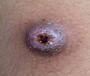
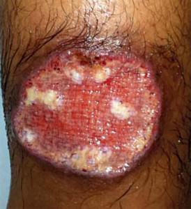
The first sign of the localized cutaneous form is the appearance of an erythematous spot at the site of a mosquito bite, after an incubation period ranging from 2 weeks to 3 months.9 This spot evolves into a papule, that progressively ulcerates over a period of 2 weeks to 6 months (Figures 1 and 2).4 The typical lesion is a round or oval painless ulcer, located on exposed skin areas.16 Although the ulcerated form comprises the vast majority of ATL cases, the disease can evolve with indeterminate clinical features.17
FIGURE 1 Initial clinical lesion, with one – month evolution, in a case of localized cutaneous leishmaniasis. Patient evolved with ulceration and development of a typical ATL cutaneous lesion
FIGURE 2 ATL ulcer with elevated borders: evolution of the case after five months.
Exceptionally, other types of localized cutaneous lesions may be found. It is important to mention the verrucous lesions that make differential diagnosis with deep mycoses and mycobacterioses, classically referred to by the acronym PLSCT (paracoccidioidomycosis, leishmaniasis, sporotrichosis, chromomycosis and tuberculosis). Another important example is the disease form that evolves with lymphangitis, combined or not with primary cutaneous lesions, a presentation hardly differentiated from sporotrichosis by clinical examination.
Leishmaniasis recidiva cutis is yet another manifestation of localized cutaneous leishmaniasis. It is characterized by the appearance of papules, usually on the periphery of an already healed ulcer. This form tends to appear two years after the clinical cure of ATL following poor treatment or lack thereof.18 The fact that it can be considered a differentiated clinical form of ATL is controversial, but such lesions as described above deserve special attention, indicating a possible treatment resistance.
Disseminated Cutaneous Leishmaniasis
It is a rare form of ATL characterized by the appearance of at least 10 lesions, which may be papular, disseminated acneiform, ulcerated, with a high incidence of mucosal involvement.19 It is believed that, after the appearance of the primary lesion, an hematogenic or lymphatic dissemination occurs in patients with relative cellular immunodeficiency against Leishmania.20 This presentation may be accompanied by systemic manifestations such as fever, malaise, myalgia and weight loss.19
Diffuse Cutaneous Leishmaniasis
The diffuse form of ATL occurs in patients with impaired cellular immune response, necessary to fight the infection. In Brazil, it is caused by L. (L.) amazonensis.21 This clinical condition is characterized by nodular or plaque lesions, rarely ulcerated, extensive and diffuse.22 Affected patients present Montenegro skin test (MST), in general, with negative or weak reactions.23
Mucosal Leishmaniasis
The mucosal form tends to affect individuals with improperly treated cutaneous form, preceded by hematogenic dissemination of the parasite. Other factors may also be associated with mucosal involvement such as the host’s immunity, male gender, extensive primary lesions or multiple lesions above the pelvic waist, primary lesion persisting over a year and malnutrition.24
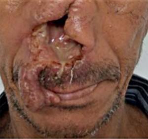
It is characterized by mucosal infiltration, which may be associated to deformities and destruction of facial structures, especially the nasal septum, and more rarely the oral mucosa or oropharynx.4,25 ( Figure 3) It represents a form of ATL that is difficult to diagnose, especially because of the challenge of obtaining biological samples for laboratory tests. The mucosal form is preferably due to L. (V.) braziliensis, although L. (L.) amazonensis may, rarely, represent the causal agent.26
FIGURE 3 Case of cutaneous leishmaniasis with 10 years of evolution. An intense destruction of the nasal septum can be observed. This case represents a severe unusual clinical lesion, however it illustrates the potential severity of mucosal leishmaniasis.
Source: scielo.br


We have not yet begun to feel the effects of all these illegal aliens. I’m sure there is rampant disease, we already know they have no respect for human life, rape, murder, pillage and go on their way. This is what is going to get worse until we send them all packing.
The mark is the symbol Islam wears on the foreheads or arm. Which in Arabic letter is a 666!
http://youtu.be/8tY3ORo7AnM
Deport them all!
I believe you are correct!
Oh goody.